

The human spine contains 33 vertebrae that work together to support your body. These remarkable building blocks of our backbone help us stand upright and create a protective tunnel for our spinal cord.
Our spine’s vertebrae are arranged into distinct sections: 7 cervical, 12 thoracic, 5 lumbar, 5 sacral, and 4 coccygeal vertebrae. Each vertebra has a cylindrical body that bears weight and absorbs shock through cushioning disks. Strong ligaments hold these vertebrae firmly in place, which allows us to bend and twist while maintaining spine stability.
In this piece, we will explore the vertebrae’s fascinating anatomy, their development, common problems that affect them, and modern diagnostic methods that keep our spines healthy.
Basic Structure of Vertebrae
The vertebral column creates a remarkable structural framework, and each vertebra serves specific functions. The vertebral column splits into five distinct regions: cervical, thoracic, lumbar, sacral, and coccyx.
Types of Vertebrae in the Human Spine
The human spine features specialized vertebrae in each region. The cervical region has seven vertebrae (C1-C7), where C1 (atlas) and C2 (axis) enable head movement. The thoracic region’s twelve vertebrae (T1-T12) connect to the ribcage to provide stability. The lumbar region has five vertebrae (L1-L5) with the largest vertebral bodies that support upper body weight. The sacrum has five fused vertebrae (S1-S5), and the coccyx has four fused bones.
Key Components of Each Vertebra
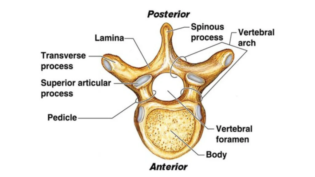
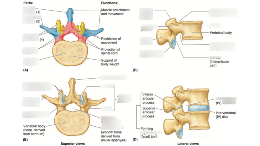
Each vertebra’s structure consists of two main parts: the vertebral body and the vertebral arch. The vertebral body sits anteriorly, serving as the primary weight-bearing component. Positioned posteriorly, the vertebral arch forms a protective ring around the spinal cord.
The vertebral arch has several vital components. Two pedicles extend from the vertebral body and connect to flat laminae that form the arch’s posterior portion. On top of that, it has seven processes that emerge from this structure: one spinous process projects posteriorly, two transverse processes extend laterally, and four articular processes. These processes serve as attachment points for muscles and ligaments.
The intervertebral disks lie between adjacent vertebrae and make up about 25% of the vertebral column’s length. These disks work as natural shock absorbers that allow spine flexibility while maintaining stability.
Evolution of Human Vertebrae
The amazing development of vertebrae spans millions of years. Nature shaped our modern spine through vital adaptations. Fish were the first vertebrates, from which all others evolved—including humans.
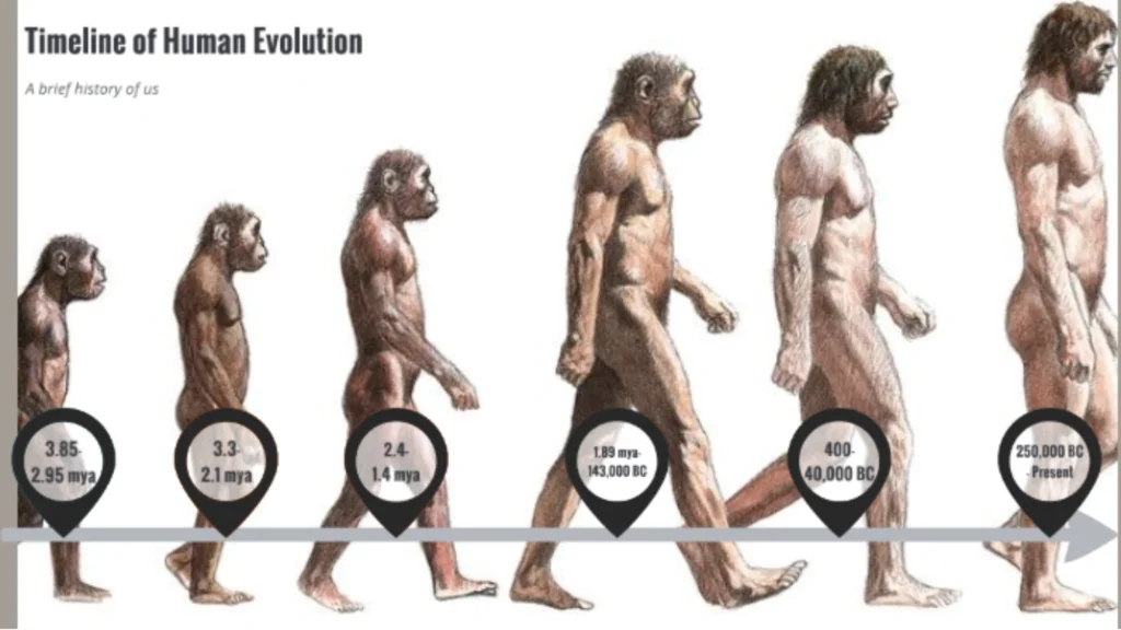
From Four-Legged to Upright Walking
Our vertebral structure changed fundamentally when we moved from quadrupedal to bipedal movement. We primarily combined apelike and humanlike ways of moving around from at least 6 to 3 million years ago. Fossil evidence shows a gradual shift from climbing trees to walking upright.
Modern Spine Adaptations
Our spine developed several vital adaptations for bipedal locomotion. The S-shaped curvature became a distinctive feature that contrasts with the C-shaped spine of quadrupedal primates. The lumbar region went through major changes, with humans typically having five lumbar vertebrae.
These changes in development brought both benefits and challenges. Our spine now efficiently:
- Distributes weight over the hips and legs
- Absorbs shock during walking
- Maintains balance in an upright position
- Supports internal organs
These adaptations came with certain costs. The spine’s new setup makes humans prone to back problems, especially in the lower back region. Studies show that regions of DNA prone to deletion during replication may have allowed vertebrates to adapt successfully to changing environmental conditions faster.
Future Evolution Predictions
Our vertebrae face new challenges today. Modern lifestyle factors, like prolonged sitting and reduced physical activity, put different stresses on our spine. Future changes might focus on addressing these modern challenges, though such adaptations would take thousands of generations.
The development of our vertebrae shows a delicate balance between improved mobility and structural stability. Understanding these changes becomes more vital as we adapt to modern life and maintain spinal health.
Common Vertebrae Problems
Spinal fractures affect more than 1.5 million Americans annually, and they range from mild to severe cases.
Vertebrae Fractures: Causes and Effects
We identified four main types of vertebral fractures:
- Compression fractures – these affect the front of vertebrae
- Burst fractures – these come with multiple vertebral breaks
- Flexion-distraction fractures – sudden forward motion causes these
- Fracture-dislocations – these create unstable vertebral movement
Almost half of new spinal injuries each year happen because of motor vehicle accidents. Osteoporosis plays another major role, particularly in women over 50. These women face a 40% risk of getting compression fractures related to osteoporosis.
C6 Vertebrae Injuries
The C6 vertebra sits near the neck’s base, and damage here affects body functions below the top of the ribcage. People who have C6 injuries can usually move their hands and arms somewhat, though they face restrictions. Their breathing control might suffer because their diaphragm’s function decreases.
Thoracic Vertebrae Issues
The thoracic spine contains 12 vertebral bodies and faces specific challenges. Disk degeneration in this area becomes the biggest problem and causes pain in the upper or mid-back. Severe degeneration can lead to bone spurs that limit how well the thoracic spine moves.
Thoracic compression fractures create another serious challenge, usually stemming from osteoporosis or trauma. Patients might suddenly feel severe back pain and become shorter. Less than 1% of all disk herniation cases involve thoracic issues, and doctors typically diagnose these in patients between 30-40 years old.
Modern Diagnosis Methods
Medical imaging has seen remarkable progress that changed how doctors diagnose and treat vertebrae conditions. The combination of aging populations and better access to advanced imaging has led to a dramatic rise in spine-related imaging over recent decades.
3D Imaging Technologies
We moved from 2D to 3D imaging because of breakthroughs in CT scan slice thickness. Today’s 3D imaging gives doctors an unmatched view of spinal structures. Modern systems can capture full-body, high-quality images in seconds. These advanced systems reduce radiation exposure by up to 80% compared to traditional methods.
Modern 3D imaging technologies deliver several key benefits at once:
- Creation of detailed surgical planning models
- Better visualization of complex spinal anatomy
- Precise measurement of spinal parameters
- Better assessment of tumor encroachment on vital structures
AI-Powered Spine Analysis
AI has transformed spine diagnosis through machine learning models. These systems now achieve diagnostic accuracies of 83-88% for spinal canal analysis and 71-75% for lateral recess evaluation on axial CT scans.
AI integration has cut diagnosis time from 10 minutes to just 14.5 seconds, matching experienced doctors’ accuracy at 98%. These AI systems excel at vertebral fracture detection, stenosis identification, and tumor detection.
Advanced MRI techniques combined with AI algorithms enable faster scan times through deep learning de-noising techniques. New specialized software can automatically generate quantitative spinal parameters such as T4-T12 kyphosis and pelvic tilt from radiographs.
Conclusion
The human vertebrae give us an amazing explanation of anatomy and medical progress. Our research shows how 33 vertebrae collaborate in distinct sections. They enable movement and protect our spinal cord.
The spine’s development tells a remarkable story. It changed from a basic structure in fish to a complex S-shaped column that lets humans walk on two legs. These changes brought many advantages to humans, but they also made our spines prone to various conditions.
Medical science moves faster every day, especially when it comes to vertebrae diagnosis and treatment. New 3D imaging technologies combined with AI analysis help doctors detect spinal problems with better accuracy and speed. These improvements lead to better patient outcomes and reduce radiation exposure and diagnosis time.
Our understanding of vertebrae continues to grow. Dedicated scientists and medical professionals develop new ways to understand and treat spinal conditions. We have a long way to go, but we can build on this progress. Their work brings hope to millions of people affected by vertebral injuries and disorders, and promises better solutions for future generations.